Hoof, Body & Soul, Part III: Mission Impossible, unedited, by Gudrun Buchhofer. Blog 62, case # 62
CHAPTER 1
Atrophy, the fundamental cause for most all hoof pathology and upper body injuries
Black hole syndrome; hematoma or chronic abscessing
Case # 62
Pete
trimmed from 2012 until 2018
July 2012
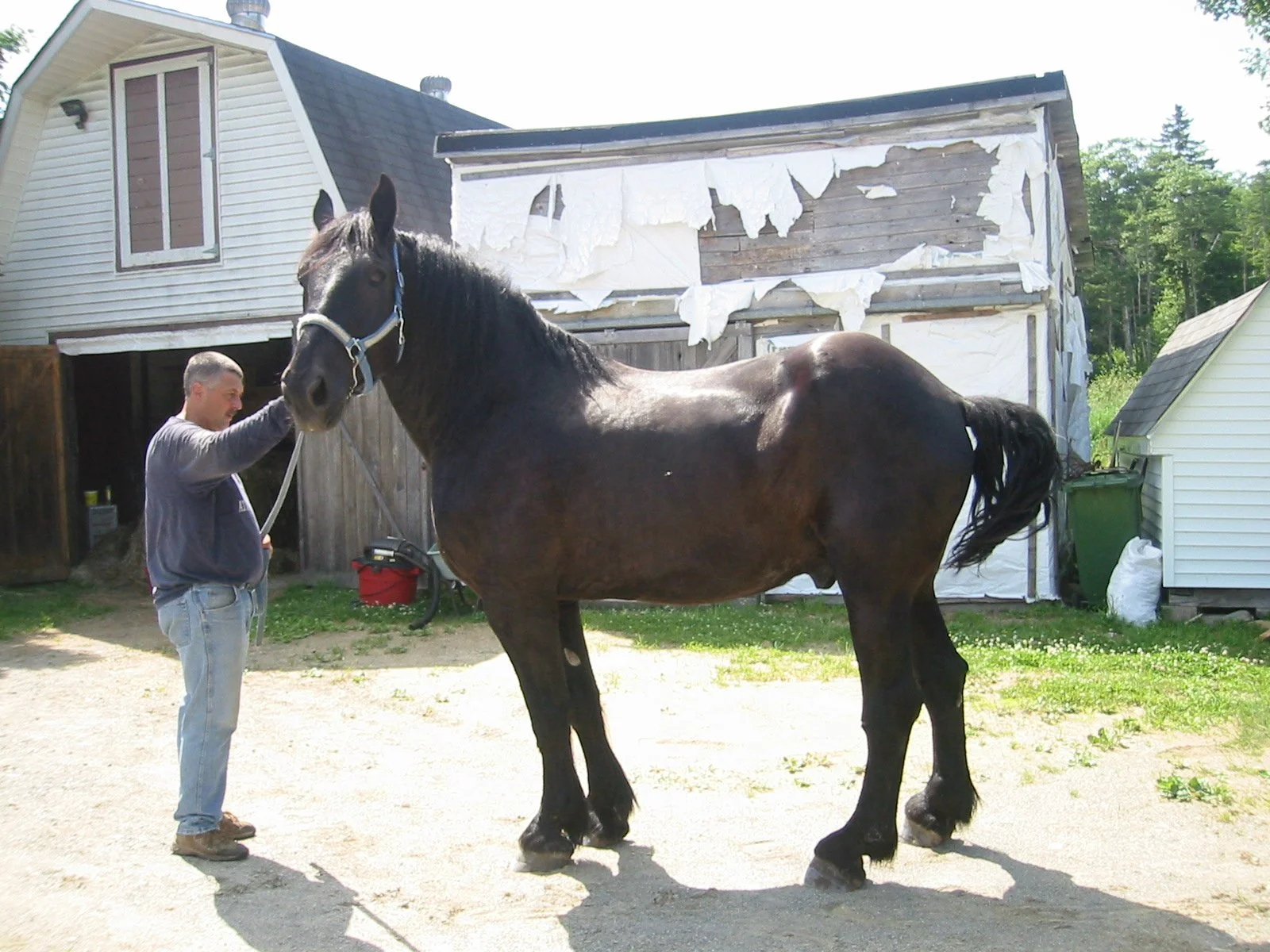
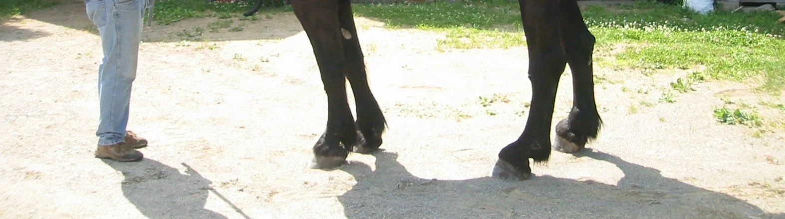
Pete was under my hoof care wings from the age of fourteen for the last six years of his life. When I first met him, the black Percheron was standing with the front legs tucked behind the vertical (toeing out with the right front) and holding his head high. He turned out the right hind leg and braced his right hind hoof on the lateral toe and side. Even with the shoe on he avoided to put weight on the medial heel.
All four hooves were atrophied in the back. They did not support the bone column of the legs. The upper body was dealing with the consequences. Pete’s hind end was very weak. He had dropped fetlocks in both hinds.
June 2014
May 2015
The previous farrier kept a shoe on Pete's right hind to prevent him from being lame. Nevertheless, there were spouts of lameness and abscessing. I was asked to take Pete out of shoes and become his trimmer.
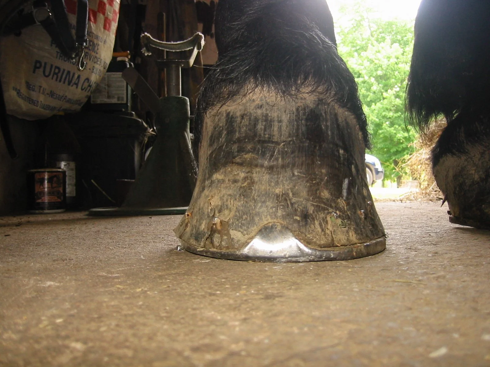
Right hind July 2012 — Pete braced on the lateral side of the right hind; he wore down the metal shoe; see the difference in thickness of the metal shoe, showing the area he put his weight on (thinning the metal)
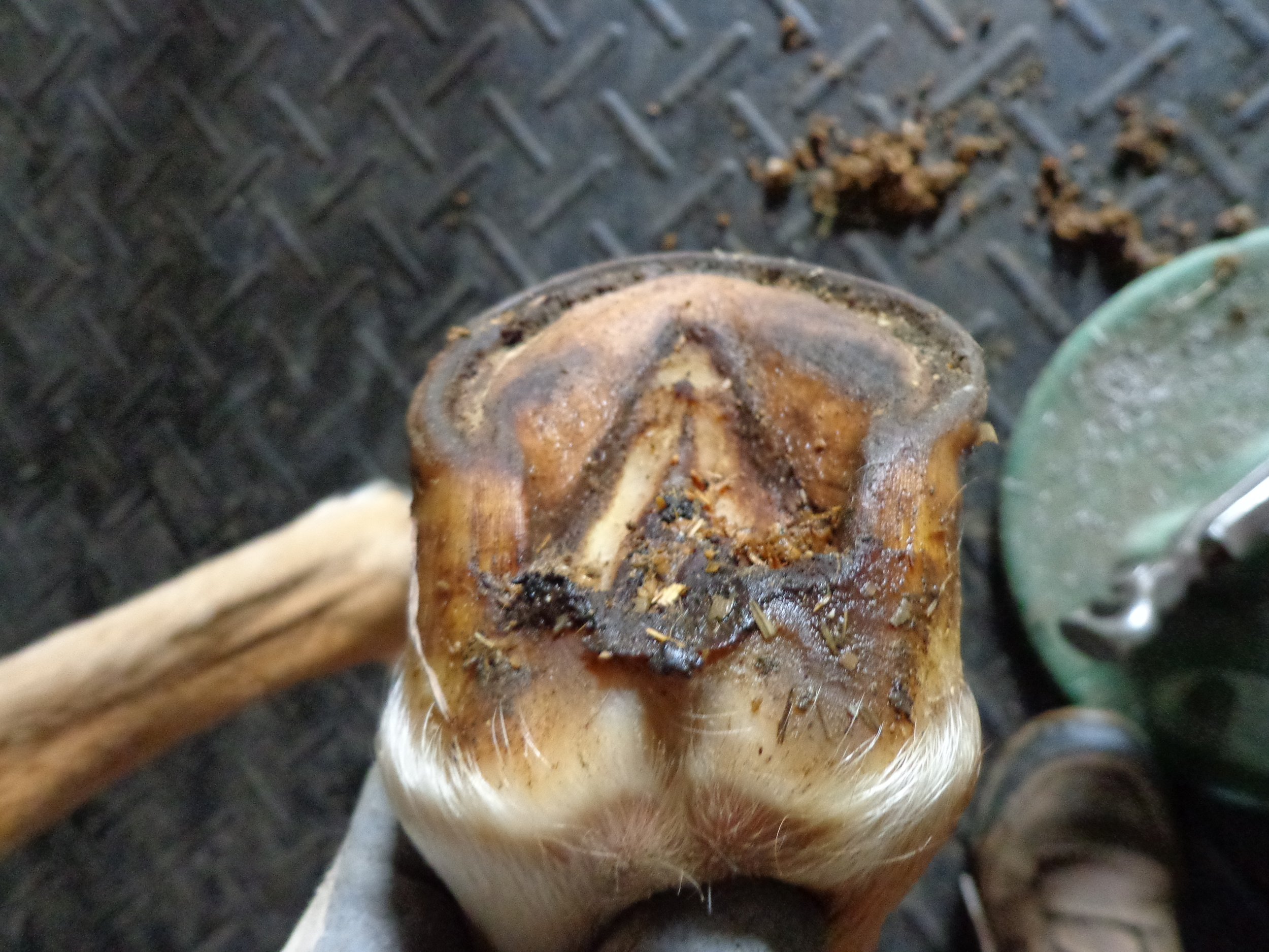
Right hind July 2012 — the picture shows the "dead corner" in the back of his foot on the medial side; despite of the metal shoe nailed on to his hoof capsule Pete did not put weight on the medial side. He leaned on the lateral side in order to avoid putting weight on the false heel. See the air gap!
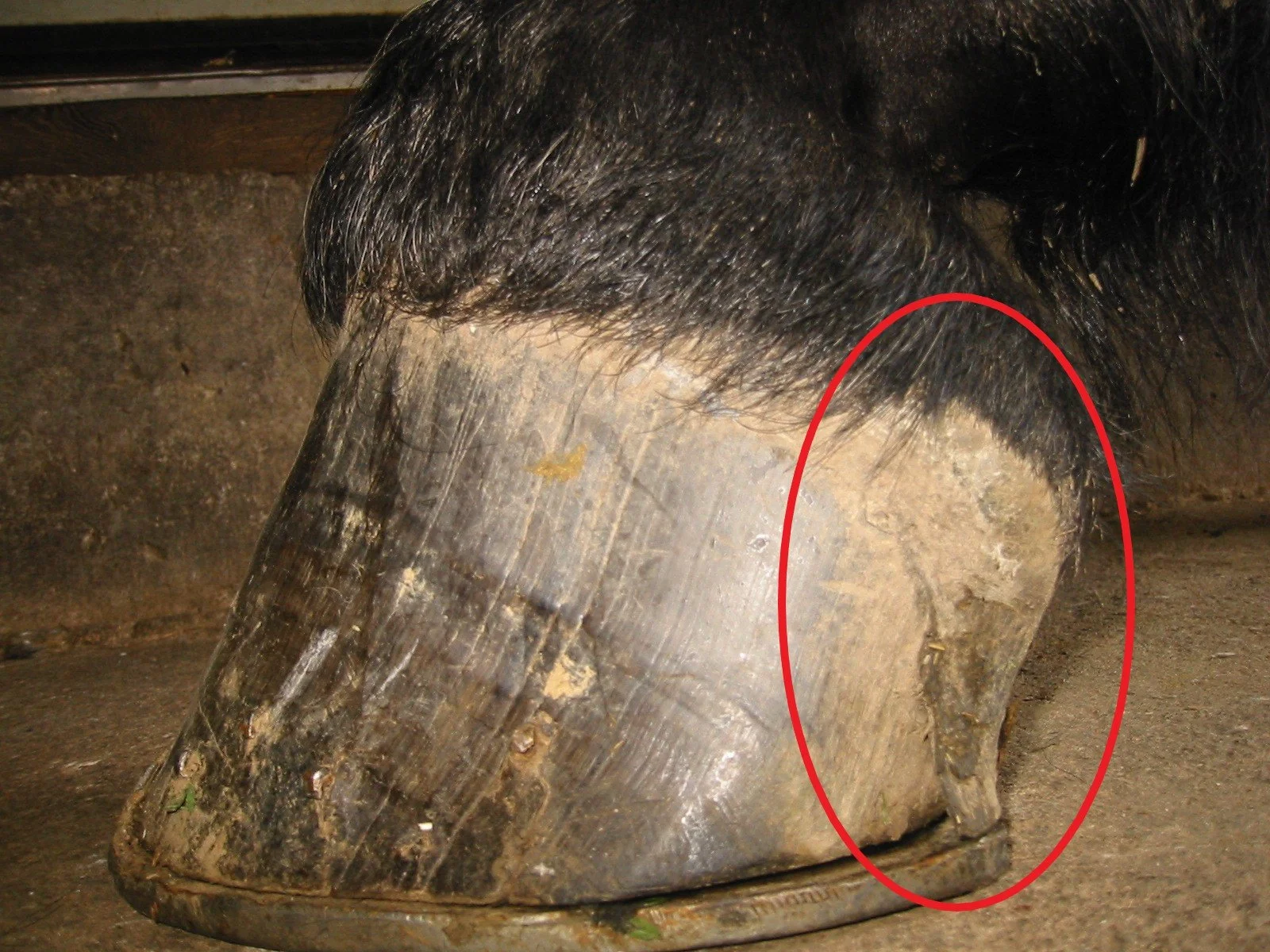
The photo of Pete’s right hind before I pulled the shoes in 2012 shows the "dead corner" in the back of his foot on the medial side. The "false heel" was meant to wear down after birth and leave his body during the first months of his life. Instead Pete was still stuck with those long shanks. The tips of the false heels had flared and grown in the soft tissue. (It looked like the claw of an eagle pinching in to the soft horn.) The variant capsule angle-of-growth in the back of the foot did not finalize after birth.
The chronic abscess channel on the right hind hoof was off to the side. It was where Pete braced himself and put on the biggest pressure in order to be able to stand. Despite the fact that Pete had four black hooves the horn along the abscess channel turned white. The hair above the cornet band turned white as well.
November 2016 — the chronic abscess channel on the right hind hoof was off to the side; the green line is marking the centre of the foot.
Right hind February 2017
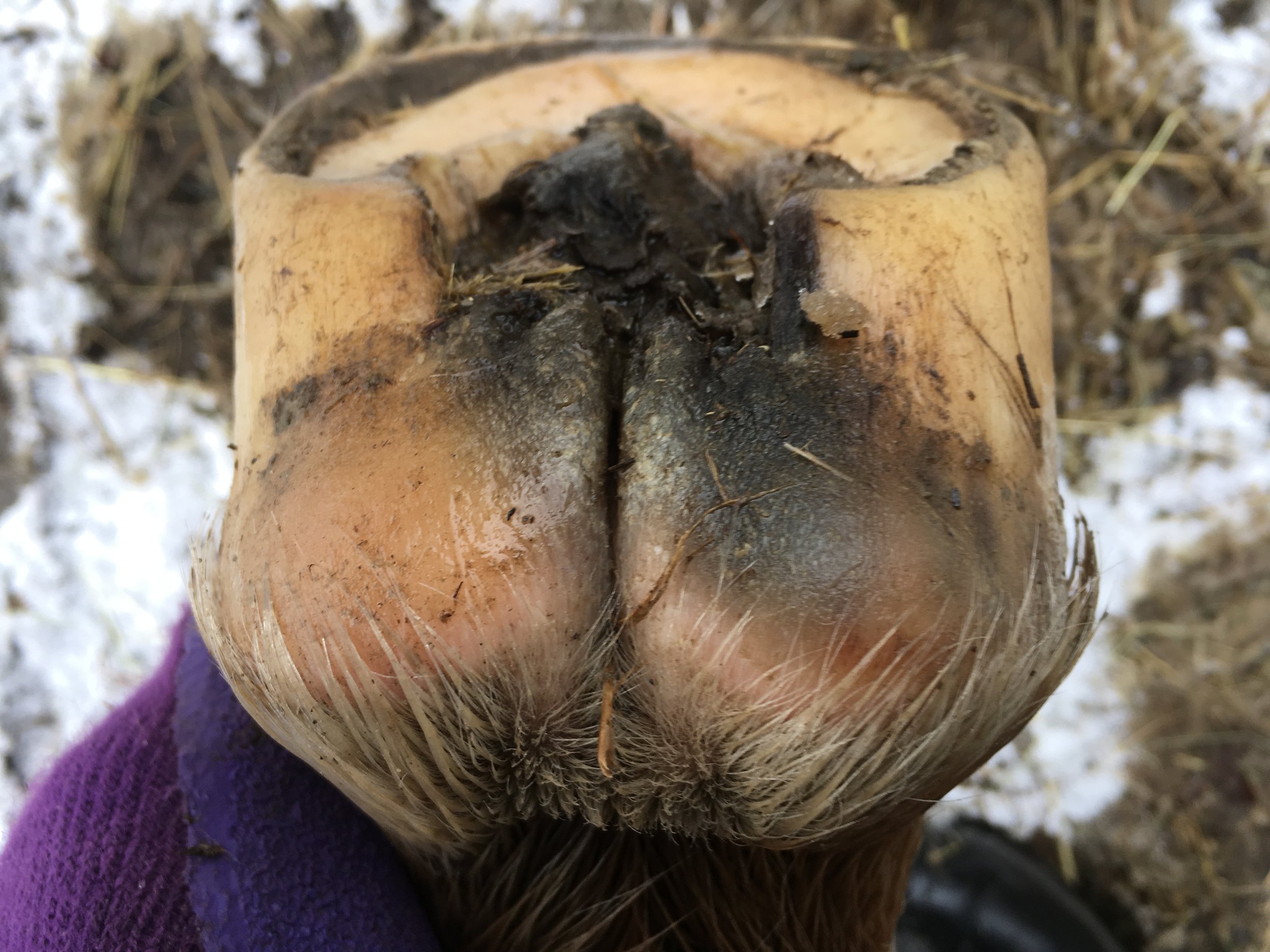
Right hind July 2017 — five years after my first trim Pete released the long shank (or false heel) from the right hind
Right hind August 2017
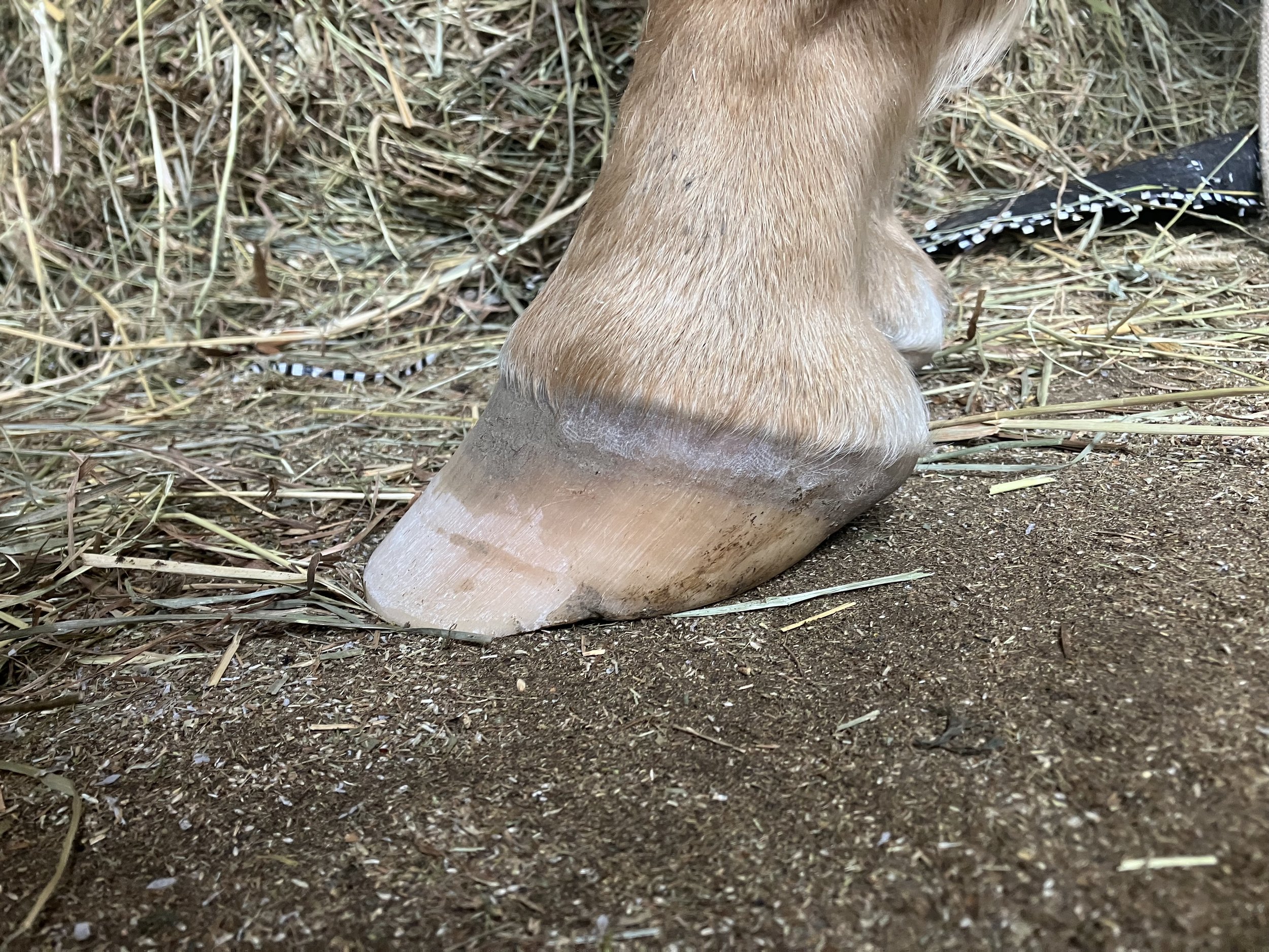
Right hind February 2018 — hoof horn had shifted around the coffin bone; the entire medial side restructured
Pete abscessed the entire frog multiple times as well as the sole of his right hind to release bruised tissue. The long shanks, or false high heels, came down over time. Hoof horn had shifted around the coffin bone. In July 2017, five years after my first trim, Pete released the long shank (or false heel) from the right hind. Although the tip of the heel was still flared and ingrown, the finalization of the variant capsule angle-of-growth in the back was in process.
Right hind February 2017
Right hind August 2017 — although the tip of the heel was still flared and ingrown, the final healing of the back of the foot was in process
All four hooves were equally involved in the pathology and the healing from it.
Right front medial side August 2017 — the previously underrun false heels are straightening up (preparing to exchange with true heels)
Right front medial side August 2017 — the previously underrun false heels are straightening up (preparing to exchange with true heels)
Right front August 2017 — the previously underrun false heels are straightening up (preparing to exchange with true heels)
July 2012 after my first trim
August 2017 — the left hind shows the increase of internal structures in the back of the foot and how much more the entire hoof capsule is under the bone column
Sadly, we run out of time. Shortly after his life long Percheron friend died, Pete passed suddenly at the age of twenty. Pete and Darky did everything together as a team. ♥♥
photos: Gudrun Buchhofer
Stay tuned for the upcoming cases (under my care for up to 20 years) in this blog series as a replacement for the unpublished part III: Mission Impossible of my trilogy Hoof, Body & Soul.
What did all my client horses over the last 20+ years have in common? They needed to heal from atrophy of the back of the foot as well as other atrophied hoof structures.
Q: Why do we need to change the upbringing of our baby horses and donkeys? A: To prevent senseless suffering.
Gudrun Buchhofer